
Lung damage patterns on CT scans may differ by ANCA, AAV types
Pulmonary involvement can help predict prognosis: Small study
Written by |

People with ANCA-associated vasculitis (AAV) may present different lung involvement patterns depending on the type of disease and self-reactive antibodies driving the disease, according to a small study.
Given that “the type of pulmonary involvement in AAV is important to predict prognosis and mortality as well as for treatment decisions by physicians,” the researchers wrote, these findings may help improve care for AAV patients.
The study, “Which is important in thoracic CT findings: Antineutrophil cytoplasmic antibody (ANCA) subtype or subgroups of ANCA-associated vasculitis?” was published as a letter in the International Journal of Rheumatic Diseases.
AAV is usually caused by anti-neutrophil cytoplasmic autoantibodies, ANCAs. The most common targets of these self-reactive antibodies are myeloperoxidase (MPO) and proteinase 3 (PR3), two proteins found in a type of white blood cells called neutrophils.
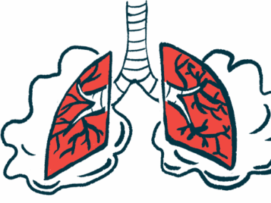
When ANCAs attach to their target proteins, neutrophils go haywire, leading to inflammation in small blood vessels and damage to several tissues and organs, most commonly the kidneys and lungs.
Microscopic polyangiitis (MPA), the most common AAV type, is usually accompanied by MPO-ANCAs. Anti-PR3 antibodies are mostly found in people with another common AAV type called granulomatosis with polyangiitis (GPA). PR3-ANCAs and GPA both have been linked to a higher risk of relapse.

Patterns of lung involvement in 64 adults with AAV examined
Now, a team of researchers in Turkey evaluated whether ANCA and AAV types influenced the patterns of pulmonary involvement in 64 adults with AAV who had abnormal findings in high-resolution thoracic (chest) CT scans.
Patients, 70.3% of which were men, received their diagnosis of AAV at a median age of 56. Nearly two-thirds (62.5%) had GPA, while the remaining 24 (37.5%) had MPA.
Among those with GPA, most (77.5%) tested positive for anti-PR3 antibodies, five (12.5%) for MPO-ANCAs, and three (10%) tested negative for ANCAs. More than half of MPA patients (58.3%) had anti-MPO antibodies, two (8.3%) had PR3-ANCAs, and eight (33.3%) didn’t have such antibodies.
When the researchers analyzed patients based on their ANCA type, they found that several lung damage patterns were significantly more common in those with MPO-ANCAs than in those positive for anti-PR3 antibodies.
The features included interlobular septal thickening (55.6% vs. 18.2%), bronchiectasis (52.6% vs. 18.2%), reticulation (44.4% vs. 9.1%), honeycombing (36.8% vs. 3.1%), and mosaic attenuation (21.1% vs. 0%).
Interlobular septal thickening refers to thickening of the walls that separate different parts of the lungs. Bronchiectasis occurs when the airways become damaged and widened, forming pouches that make it hard to clear mucus from the lungs.
Reticulation and honeycombing, with or without bronchiectasis, are features of usual interstitial pneumonia, a pattern of interstitial lung diseases, or diseases that cause the lungs to scar.
Mosaic attenuation refers to patchwork of regions of differing attenuation on lung CT imaging that may reflect vascular disease and airway abnormalities.
When patients were analyzed according to their AAV type, the team found that a significantly higher proportion of those with MPA showed interlobular septal thickening (47.8% vs. 17.5%), reticulation (34.8% vs. 10%), and honeycombing (29.2% vs. 5.1%) relative to those with GPA. Airway bleeding also was significantly more common in the MPA group (33.3% vs. 7.5%).
In turn, a significantly higher proportion of GPA patients showed nodules (62.5% vs. 8.3%) and cavities (37.5% vs. 0%) in their lung CT scans, when compared with those with MPA.
“Classification of patients according to ANCA subtype shows that patients with MPO-ANCA may have high-resolution CT findings related to interstitial pneumonia,” the researchers wrote.
Similarly, people with MPA, a type most commonly associated with anti-MPO antibodies, “had more frequent CT findings related to interstitial pneumonia, as well as [airway bleeding],” they added.


Leave a comment
Fill in the required fields to post. Your email address will not be published.