CT scan damage may predict ANCA-associated vasculitis outcomes
Certain lung damage patterns associated with higher relapse rates
Written by |
Patterns of lung damage on CT scans may help predict disease outcomes in people with ANCA-associated vasculitis (AAV) and determine which patients should receive more intensive treatment, a study in Germany suggests.
Particularly, ground glass opacities (GGO), which are white diffuse regions due to fluid, airway collapse, tissue scarring, and others, and fluid buildup in the membrane that surrounds the lungs, called pleural effusion, were each associated with higher relapse rates. Lung nodules were linked to lower rates of relapses.
“Our data demonstrate the outstanding importance of characteristic CT patterns in AAV diagnosis assessment,” wrote the researchers, who said “certain CT patterns including GGO and pleural effusion may help to identify patients who are at higher risk for relapsing disease.”
The study, “Distinct pulmonary patterns in ANCA-associated vasculitides: insights from a retrospective single center cohort study,” was published in Rheumatology International.
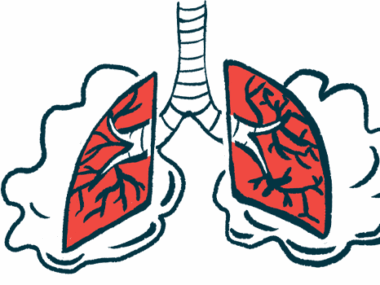
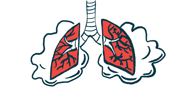
AAV is a group of autoimmune diseases marked by inflammation and damage to small blood vessels that are typically caused by self-reactive antibodies called ANCAs. While AAV types may involve different organs and tissues, “lung involvement is one of the most prevalent clinical features in [AAV] and significantly contributes to disease burden and mortality in these patients. Reported rates of pulmonary affection in the two major AAV [types] range up to 85% in GPA [granulomatosis with polyangiitis] and up to 50% in MPA [microscopic polyangiitis] patients,” the researchers wrote.

Analyzing CT scans to predict outcomes
Researchers at the Aachen University Hospital retrospectively analyzed the clinical data and chest CT scans of 94 AAV patients treated at their hospital between 1999-2020.
CT scans were screened for predefined patterns, including ground-glass opacities, presence of micronodules (smaller than 3 mm, or about 0.11 inches), nodules (3 mm to 3 cm, or about 1.18 inches), and masses (bigger than 3 cm), cavities (collection of gas and/or fluid enclosed by a thick wall), interstitial lung disease (ILD), pleural effusion, and lymphadenopathy, or swollen lymph nodes in the chest. ILD comprises a group of conditions marked by tissue scarring.
The patients’ mean age was 56.4 and 54.3% were males. More than half (52.1%) had GPA, 43.6% had MPA, and the AAV type wasn’t able to be determined in the remaining four patients (4.3%). Lung involvement was present in 44 patients (46.8%) with available CT scan data. Most had GPA (70.5%), and kidney involvement (72.7%).
Over a median follow-up of about three years, patients with lung involvement were 71% more likely to relapse and 82% more likely to die, but these differences weren’t statistically significant. These patients were also nearly six times more likely to undergo plasma exchange as an add-on to standard immunosuppressive treatments. Plasma exchange involves removing and replacing plasma, the liquid portion of blood that contains antibodies, including ANCAs.
Among the patients with lung involvement, rituximab (sold as Rituxan, among others) and cyclophosphamide were used to induce disease remission in a similar proportion, about 36% each. “Both treatment options did not differ significantly regarding mortality… or relapse rate,” the researchers wrote.
When comparing lung damage patterns between MPA and GPA patients, a significantly greater proportion of those with MPA had ground glass opacities (72.7% vs. 19.4%) and interstitial lung disease (72.7% vs. 32.3%). Lung nodules were significantly more common among GPA patients (64.5% vs. 27.3%).
Pleural effusions were more frequent in the MPA group (54.5% vs. 22.6%), lung cavities were more common among GPA patients (19.4% vs. 9.1%), and masses were exclusively detected in this latter group (25.8% vs. 0%). These differences didn’t reach statistical significance, however.
Lung opacities were significantly associated with a nearly nine times higher likelihood of relapse and pleural effusions with a six times higher chance of relapse. Patients with nodules were 85% less likely to relapse.
The presence of cavities was significantly linked to an 89% lower chance of serious infections and patients with lymphadenopathy were seven times more likely to die. A higher mortality rate was seen among those with GPA (14.3% vs. 4.9%), but this difference didn’t reach statistical significance.
This study “identifies important subgroups such as MPA patients with signs of GGO and/or pleural effusions that are more prone to develop relapses, advocating for a more severe immunosuppressive regimen upon initial diagnosis,” the researchers wrote. “If our observations are confirmed in larger, multicentric [patient groups], future therapeutic trials should prospectively evaluate more individualized immunosuppressive regimens in these subsets of AAV patients.”