
Blood Markers May Help Distinguish Lung Infection From AAV Flares
Elevated PCT levels in patients' blood signal infection, affecting treatment
Written by |
Higher blood levels of the molecule pro-calcitonin (PCT) are a marker of lung infection among ANCA-associated vasculitis (AAV) patients with lung disease, a new study reports.
Combining PCT with other blood markers can further increase the accuracy of identifying infection, and may be useful for distinguishing between AAV-related lung involvement and lung infection in AAV patients, which has important implications for clinical care.
The study, “Clinical features and markers to identify pulmonary lesions caused by infection or vasculitis in AAV patients,” was published in BMC Pulmonary Medicine.

Symptoms of lung infection and injury similar in ANCA-associated vasculitis
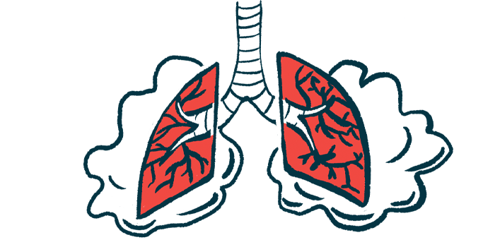
AAV is characterized by inflammation in the blood vessels that can damage organs, including the lungs. AAV lung involvement can result in symptoms like fatigue and cough, similar to what you might expect from an infection in the airways.
Imaging scans of the lungs can detect AAV-associated injury, but it is often similar to damage due to an infection.
The similarity between AAV-related lung damage and lung infection can be a diagnostic challenge for clinicians, especially since people with AAV often have weakened immune systems and are at increased risk of infections.
When AAV patients start to show signs of new lung problems, it’s critical to determine whether the cause is an AAV flare or an infection.
“Accurately identifying the nature of lung injury in patients with AAV is very important because unreasonable treatment based on the wrong diagnosis may lead to serious consequences,” the researchers wrote.
While giving immunosuppressing medicines can help to control AAV, they may worsen an infection by weakening the body’s ability to fight it. Conversely, inappropriately giving antibiotics or other infection-targeting treatments could mean delaying treatment for AAV flares that tend to worsen over time, the researchers noted.
A team of researchers in China conducted an analysis looking for markers that might be useful for distinguishing infection from AAV-related damage in patients with lung disease.
The analysis included data from 140 people with AAV who received care at the Renmin Hospital of Wuhan University between 2016 and 2021. Among these patients, 118 had signs of lung disease, or lesions — specifically, 68 had lung infections, while the other 50 had no infections, so their lesions were presumably a result of AAV.
Among patients with lung disease, the researchers noted that certain symptoms were more common among those with infections.
Specifically, patients with lung infections were significantly more likely to experience fever (52.94% vs. 16%), chest tightness (61.76% vs. 40%), cough and expectoration (coughing up phlegm or mucus) (72.06% vs. 46%), and coughing up blood (27.94% vs. 8%) than those without infections. Fever duration also was significantly longer in the lung-infection group than in the non-infection group.
Certain lung imaging features, likewise, were more common among patients with infections.
These findings suggest “that complications with infection could lead to more severe clinical symptoms,” the researchers wrote.
“However,” they added, “clinical and [imaging] manifestations are not fully specific to the nature of lung injury and are insufficient to distinguish between lung infection and vasculitis injury.”
Blood levels of pro-calcitonin, or PCT, a known marker of infection
Notably, patients with lung infection had significantly higher values of several known markers of infection than patients in the other two groups.
These included higher blood levels of PCT, C-reactive protein (CRP), and amyloid A, as well as a higher blood neutrophil-to-lymphocyte ratio (NLCR) and erythrocyte sedimentation rate (ESR). NLCR is a measure of immune cell counts, and ESR measures how fast red blood cells settle inside a test tube.
The lung-infection group also had a significantly higher Birmingham vasculitis activity score, which measures AAV activity and organ involvement.
Statistical analyses then tested the markers’ ability to discriminate between AAV patients with and without lung infection.
Scientists found that the best marker was blood levels of PCT. Specifically, a cutoff of 0.235 nanograms per milliliter allowed them to correctly identify lung-infection patients with a sensitivity of 85.3% (true-positive rate) and a specificity of 84% (true-negative rate).
Combining PCT with other markers, specifically CRP and NLCR, further improved the accuracy of “distinguishing the infectious and non-infectious lung injuries in AAV patients,” the researchers wrote.
“The composite indicator of PCT-CRP-NLCR had the highest diagnostic value,” the team concluded, suggesting that this combination could be used to help identify lung infection among AAV patients with lung disease.


Leave a comment
Fill in the required fields to post. Your email address will not be published.