Japanese Case Study Links Patient’s Microscopic Polyangiitis to Silica Dust
Written by |

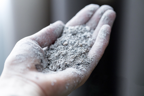
Prolonged exposure to the silica dust that construction and other industries generate contributes to the development of the small blood vessel disease known as microscopic polyangiitis, according to a Japanese case study.
Microscopic polyangiitis, or MPA, is one of three types of a category of diseases known as ANCA-associated vasculitis, or AAV. The conditions’ hallmarks are inflammation in, and destruction of, small blood vessels. The other two types are granulomatosis with polyangiitis and eosinophilic granulomatosis with polyangiitis.
Scientists don’t fully understand what causes MPA, but they know that factors like drugs, bacteria, and exposure to environmental hazards are important contributors.
The autoantibodies associated with the disease can strike small vessels in different organs. Patients often experience kidney, lung, or neurological symptoms. An autoantibody is a protein the immune system generates against another protein in the body rather than an invader.
Researchers at Ako Central Hospital, in Hyogo, Japan, reported the case of a patient with prolonged occupational dust exposure who was diagnosed with MPA. The study in the Journal of General and Family Medicine is titled “Silicosis, then microscopic polyangiitis—antineutrophil cytoplasmic antibodies‐associated vasculitis may be work‐related disease in patients with silicosis.”
The 74-year-old retired shipyard worker went to a hospital for fatigue and two weeks of persistent fever. He also reported a dry cough, but no additional symptoms or weight loss.
Doctors had previously diagnosed him with silicosis, a lung disease stemming from long-term exposure to silica dust. The condition is characterized by inflammation and scaring of lung tissue. The patient had worked in a shipyard — a profession that exposed him to the dust — for 43 years until his retirement.
In addition to the fever, he had crackling sounds in his lungs and a rapid breathing rate. Laboratory work showed he also had blood in his urine.
A computed tomography scan showed a dark area in his lungs that led to an initial diagnosis of pneumonia. Doctors gave him an oral antibiotic for a week, but he failed to improve.
Because he continued to experience fever and kidney dysfunction, doctors did additional blood tests. These revealed high levels of autoantibodies against the protein myeloperoxidase, or MPO. This prompted doctors to diagnose him with MPA and a rapidly progressing kidney disease.
Doctors ordered immunosuppressive therapies to counter his MPA. They also instituted aggressive treatment regimens to manage his kidney symptoms. The patient “was discharged from the hospital approximately four months after admission, following improvement of his symptoms,” the researchers wrote.
Scientists have speculated “that particles in environmental silica induce systemic autoimmunity,” the team wrote. “In our present case, the patient developed MPA following 43 years of occupational silica exposure.”
Silica exposure alone could not explain the onset of AAV, the team noted. They said an invasive procedure used to manage his lung condition could have triggered the disease.
“In conclusion, with respect to work-related disease and potential compensation, it is essential to recognize silica exposure as a risk factor for developing AAV such as MPA,” researchers wrote. “Our case emphasizes the importance of primary physicians identifying this relationship in addition to taking a thorough occupational history.”


