
Blood Levels of Proteins Identify Active AAV in Long-term Study
Written by |

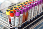
Blood levels of several proteins — CXCL13, interleukins IL-6, IL-8, IL-15, IL-18 binding protein (IL-18BP), matrix metalloproteinase-3 (MMP-3), and erythrocyte sedimentation rate (ESR) — were validated as effective at identifying active ANCA-associated vasculitis (AAV) in a long-term study.
The study, “Serum Biomarkers of Disease Activity in Longitudinal Assessment of Patients with ANCA-Associated Vasculitis,” was published in the journal ACR Open Rheumatology.
AAV is a group of autoimmune diseases characterized by inflammation and damage to small blood vessels. After the initial treatment, the disease’s progression is variable, and clinicians have a hard time distinguishing flares from symptoms caused by infections, adverse reactions to therapies, or other causes.
To predict relapses and differentiate active disease from remission, physicians often rely on the levels of ANCAs — the self-reactive antibodies that cause inflammation in AAV — as well as generic markers of inflammation such as ESR and C-reactive protein (CRP). However, these tests are not enough to determine how individual patients should be treated.
In the previous RAVE Phase 2/3 clinical trial (NCT00104299), which investigated the antibody rituximab (brand names Rituxan, Truxima, Ruxience, and others) as a remission-inducing treatment for AAV, researchers also assessed the levels of 28 blood proteins for their ability to distinguish severe AAV from remission in 137 patients.
Twenty-four of these proteins were effective at differentiating severe AAV and remission and also between active AAV and healthy participants. Three of these proteins — CXCL13, MMP-3, and TIMP-1 — were even better than than ESR or CRP at distinguishing severe AAV from remission.
Now, a team of researchers in the U.S., the Netherlands, and Iceland, supported by the Vasculitis Clinical Research Consortium (VCRC), sought to investigate 22 of the 24 proteins in samples from 74 participants in the RAVE trial, collected over a year and a half after enrollment.
Goals were to determine the ability of each protein to distinguish active AAV from remission in a setting closer to that found in clinical practice, as well as to find out how well these markers in combination could distinguish active AAV from remission. Another aim of the study was to assess if changes in protein levels during remission could predict flares.
The study included only patients who were in remission six months after starting on their assigned treatment. Blood samples were collected at screening and at 1, 2, 4, 6, 9, 12, 15, and 18 months after enrollment, as well as during flares.
Among the 74 patients, 41 were women, and their median age was 51. Most (77%) had granulomatosis with polyangiitis (GPA) and relapsing disease (52.7%); the remaining were new to the disease at trial entry.
After a year, 42 patients had experienced a relapse and 32 stayed in remission. Among patients who had relapses, the median time to first flare was 159 days (about five months) after the six-month visit. In patients with severe relapses, a total of 22, the median time to a severe flare was 205 days (almost seven months) after that six-month assessment.
The researchers used three statistical methods to evaluate the blood levels of the 22 proteins. Six of them – CXCL13, IL-6, IL-8, IL-15, IL-18BP, and MMP-3 — as well as ESR, were significantly associated with disease activity over the 18-month period in all three analyses.
Three of them, IL-8, IL-15, and IL-18BP, were the most promising markers for predicting a relapse, with a 2.7-fold increase in any of them being associated with a 40–80% greater likelihood of a relapse.
Further analysis showed that the combination of CRP, IL-18BP, neutrophil gelatinase-associated lipocalin (NGAL), and soluble IL-2 receptor (sIL-2R)-alpha could distinguish active disease from remission moderately well, with an accuracy of 72%.
Levels of CXCL13 and MMP-3 were elevated in patients taking the corticosteroid prednisone independently of disease activity, which could make their interpretation as biomarkers of active AAV difficult, according to the researchers.
The team also noted that protein concentrations during remission could not predict future flares and that markers that rise at the same time as a flare deserve further research.
“Although this study validated several markers as being associated with active AAV, the goal of finding generic markers of inflammation or ANCA titers that strongly reflect disease activity or predict future flare in AAV remains elusive,” the team wrote. “Additional hypothesis-based studies and broader agnostic screens may both be useful in pursuing these goals.”


Leave a comment
Fill in the required fields to post. Your email address will not be published.