
Rituxan’s Efficacy May Be Linked to It Blocking How B-cells and Certain T-cells Interact, Study Says
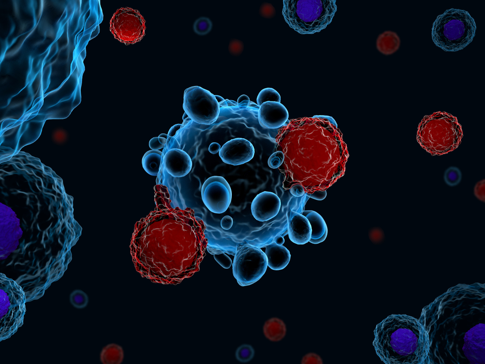


Blocking the interaction between immune B-cells and CD8-positive T-cells may be what makes Rituxan (rituximab) an effective treatment for ANCA-associated vasculitis (AAV), a study reports.
Findings of the study, “B cell depletion therapy dampens CD8+ T cell response in ANCA‐associated vasculitis,” were published in Arthritis & Rheumatology.
Anti-neutrophil cytoplasmic autoantibody (ANCA) vasculitis is an autoimmune disease caused by the production of autoantibodies — antibodies that wrongly target and attack healthy cells — leading to blood vessel inflammation and swelling in affected tissues and organs.
The disease impacts immune cells involved in both innate — general immune response against pathogens — and adaptive — specific immune response against a pathogen that involves memory — immunity, including B-cells that produce autoantibodies, CD4-positive T-cells (T-helper cells), and regulatory T-cells that regulate the function of B-cells and other types of T-cells.
Previous studies involving patients and animal models of disease suggested CD8-positive T-cells — T-cells that contain the CD8 receptor on their surface and are responsible for detecting and destroying pathogens — may also be involved in AAV. However, it is not clear how this complex auto-immune response is generated.
Two randomized clinical trials (NCT00748644; NCT00104299) demonstrated the efficacy of Rituxan in treating AAV by targeting B-cells that produced harmful autoantibodies.
Based on data that included these results, Rituxan, marketed by Genentech, is now FDA approved to both achieve and maintain remission in patients with granulomatosis with polyangiitis (GPA) and microscopic polyangiitis (MPA).
But researchers, noting that exactly how Rituxan “impacts T-cell response in AAV patients has not been fully investigated,” set out to compare the effectiveness of Rituxan and conventional immunosuppressants (CIS) — azathioprine, mycophenolate, or methotrexate — on regulatory CD4-positive, and CD8-positive T-cells in these patients.
They analyzed 63 blood samples from a total of 51 AAV patients: 20 from people with untreated active disease and 43 samples from patients in remission and either taking no treatment, or under maintenance therapy with Rituxan or CIS. The frequency of isolated T-cell sub-populations and their characteristics were assessed through in vitro assays (lab tests).
No significant differences in CD4-positive and regulatory T-cells were found between patients treated with Rituxan or conventional immunosuppressants.
But within the CD8-positive T-cell subset, Rituxan-treated patients had fewer memory effector T-cells — T-cells immediately prepared to fight a pathogen because they have a “memory” or previously encountering it — and lower production of pro-inflammatory cytokines/chemokines — molecules that mediate the immune response — compared to those treated with CIS.
These experiments also showed that CD8-positive T-cells from AAV patients, when cultured with B-cells, produced more pro-inflammatory cytokines.
But when interaction between these two types of immune cells was blocked, cytokine production dropped significantly, similar to what was seen after Rituxan treatment in AAV patients.
“We found that CD8-positive T-cells from patients in remission under CIS [conventional immunosuppressants] produced similar levels of cytokine/chemokine than those of patients with untreated active disease. Conversely, CD8-positive T-cells from patients receiving Rituxan produced lower levels of pro-inflammatory cytokine and chemokines compared to the two other groups,” the researchers wrote.
“In conclusion, the disruption of B-cell help [given] to a pathogenic CD8-positive T-cell response could contribute to the dramatic efficacy of Rituxan. Further studies are needed to determine how B-cell impact CD8-positive T-cell responses and to assess the value of CD8-positive T-cells immunomonitoring under B-cell targeted therapies,” they concluded.